
by Mr Mike Potts, Consultant Ophthalmologist BSc, PhD, FRCS, FRCOphth
As there is a rise in the medico-legal recognition of many aspects of head injury there is an increasing need for good evaluation of the levels and type of
injuries sustained both subjectively and objectively.
The visual system and its neuro-ophthalmic measurement techniques can provide evidence of BOTH subjective problems reported by the patient and the ability to support this subjective symptomatology by measurable and reproducible neuro-ophthalmic tests.
No where is it more apparent that examination of the visual system can be one of the earliest and most sensitive indicators of head injury than on the rugby field.
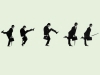
In the recent world cup matches there were numerous examples of high energy impact collisions rendering players unconscious for periods from seconds to minutes. After the player has recovered sufficiently to be orientated in space and time, the most common method that the attending doctor will use to check recovery is the player’s visual system. Both the ability to see clearly with either eye and the ability to coordinate eye movement, via the sensitive ocular motor system is commonly used by the doctors in ‘follow my finger’ routines. These routines test both acuity and coordination of eye movements.
Commonly after head injuries the after effects of concussion may last only seconds or minutes with the commonly associated stars and flashing lights. Time and a cold sponge usually helps these! Symptoms of double vision however transientary can be harbingers of more serious degrees of head injury.
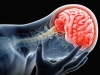
The optic nerves are the largest cranial nerves in the head comprising of 1.2 million nerve fibres in each optic nerve. Clearly long or short term damage to them is possible in head injury. Whilst short term damage will recovery rapidly, longer term damage can be easily confirmed and documented by careful measurement of visual acuity, colour vision and visual field testing. Modern automated visual field testing is extremely sensitive and reproducible. Furthermore it carries the possibility of being able to detect false positive and false negative responses being made by the patient. Hence it makes it a very reliable test which is hard to falsify. This fact is known by the DVLA and hence formal assessment of patients automated visual fields are often required by the DVLA if there is history of head injury and visual loss. Also all head injuries should follow a clear pattern of being worst initially with some slow progressive visual recovery. Repeated visual acuity testing, colour vision and automated visual field plotting will confirm this natural pattern of recovery. Patterns which are different to this will raise suspicion of their causation.
The ocular motor nerves (three, four and six) that control eye movements are also amongst the most sensitive in the brain to blunt head injury. The sixth nerve having the longest course in the head of all cranial nerves makes it especially vulnerable in blunt head trauma. Hence double vision due to sixth nerve palsies are extremely common conditions after head injury. The severity and duration of these palsies are a good indicator as to the severity of the head injury. These can be accurately and repetitively measured by orthoptic techniques which chart the severity and recovery of the double vision caused by these nerve palsies.
More fine coordination of eye movements which involve accommodation of the lens and convergence of the eye movements are also delicately balanced fine
ocular motor skills which can easily be disrupted by blunt head trauma. Their disturbance results in difficulties of fine coordination for reading and close work. This is best demonstrated in the accommodation convergent reflexes which again can be accurately and reproducibly measured by orthoptic testing.
Finally the skills of higher cortical functioning such as subtle form perception and visual memory skills are often also affected by blunt head injury. These fine
cognitive skills which can be damaged subtly in head injury are again accurately and reproducibly tested by visual and visuomotor cognitive testing often
performed either by orthoptists or by cognitive psychologists.
For all these reasons an assessment by a competent neuro-ophthalmologist of the fine visual processing skills of a patient with brain injury will often allow the
detection and measurement of the subtle types of brain injury that can result from such trauma.
Very many patients with head injury will complain of visual symptoms and their assessment and documentation is an important part of the assessment and rehabilitation of these patients. This requires a competent neuro-ophthalmologist and his supporting team of orthoptists, optometrists and psychologists.